
Microcirculation without injected dye
Mapping blood flow in the smallest vessels.
Non-invasive imaging of blood flow within the retina and choroid without an injection of dye.
Method
Non-invasive
Injection
Not required
Information
Blood-flow mapping
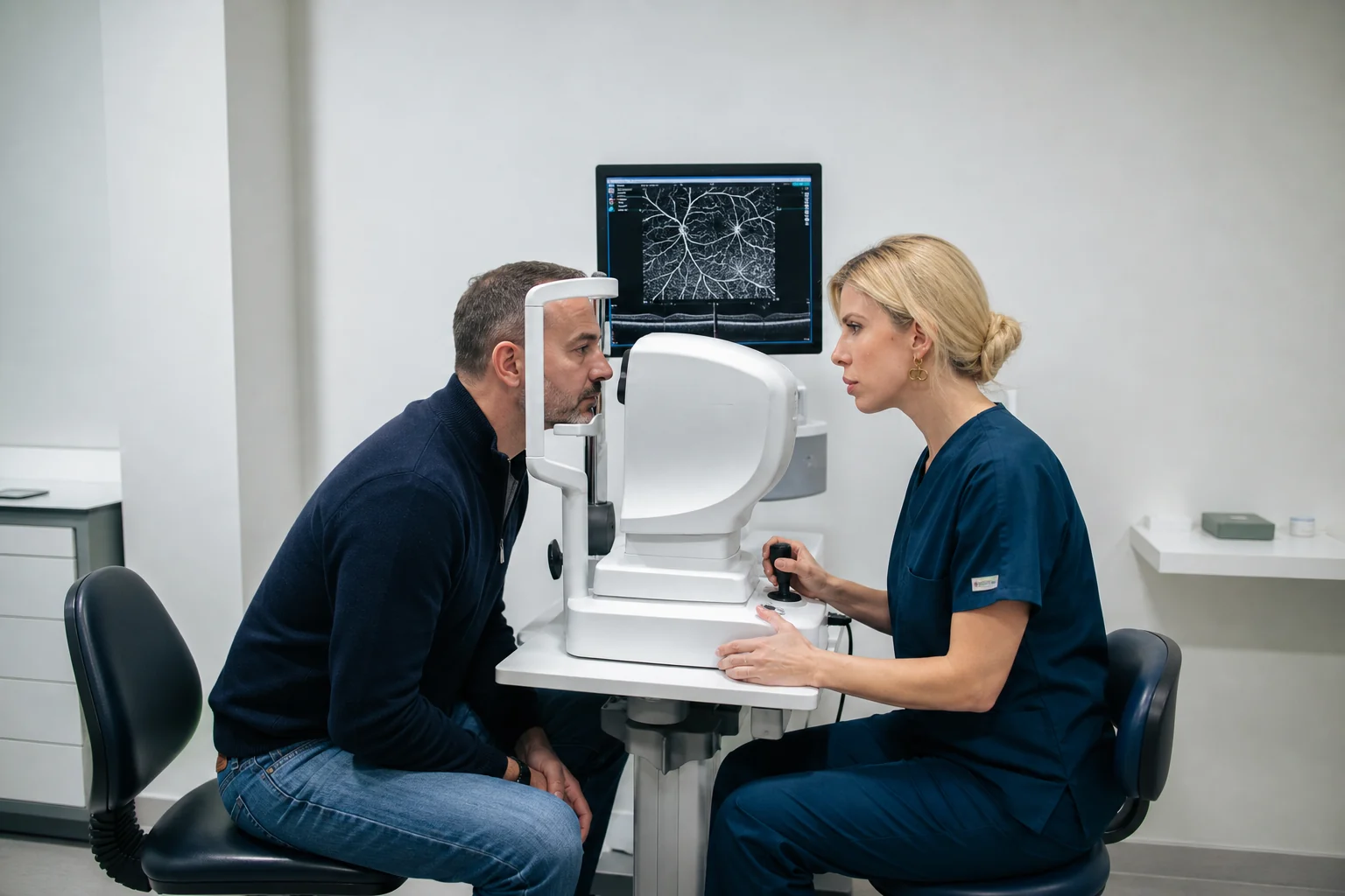
Eye examinations
OCT Angiography
What is OCTA?
A detailed view of blood flow, without intravenous dye.
OCT angiography records changes associated with blood flow in the small vessels of the retina and choroid.
It complements structural OCT and can provide information about vascular networks in selected conditions.
It is not a replacement for every form of angiography. A different examination may be required according to the clinical question.
No intravenous contrast injection is required. Image quality is affected by movement, steady fixation and any media opacity.
When it is used
A detailed view of the retinal microcirculation.
OCTA is selected when vascular information can answer a specific clinical question.
01 / MACULA
Central microcirculation
Macula
Assessment of vascular networks in conditions affecting central vision.
- Neovascular age-related macular degeneration
- Diabetic microvascular changes
- Impaired macular perfusion
- Retinal vascular occlusions
02 / FLOW
Selected indications
Vascular mapping
Complementary information for selected vascular conditions.
- Retina
- Choroid
- Selected optic nerve conditions
- Comparison with structural OCT
The process
Repeated scans without an injection of dye.
Image capture resembles conventional OCT but analyses differences associated with blood flow.
01
Positioning
The head is supported comfortably on the device.
02
Fixation
The gaze remains on a light target.
03
Flow scanning
The same area is scanned repeatedly to analyse changes.
04
Combined assessment
Findings are reviewed alongside OCT, fundus examination and other investigations.
Image limitations
Movement and unstable fixation may create artefacts, while very slow flow or leakage is not recorded in the same way.
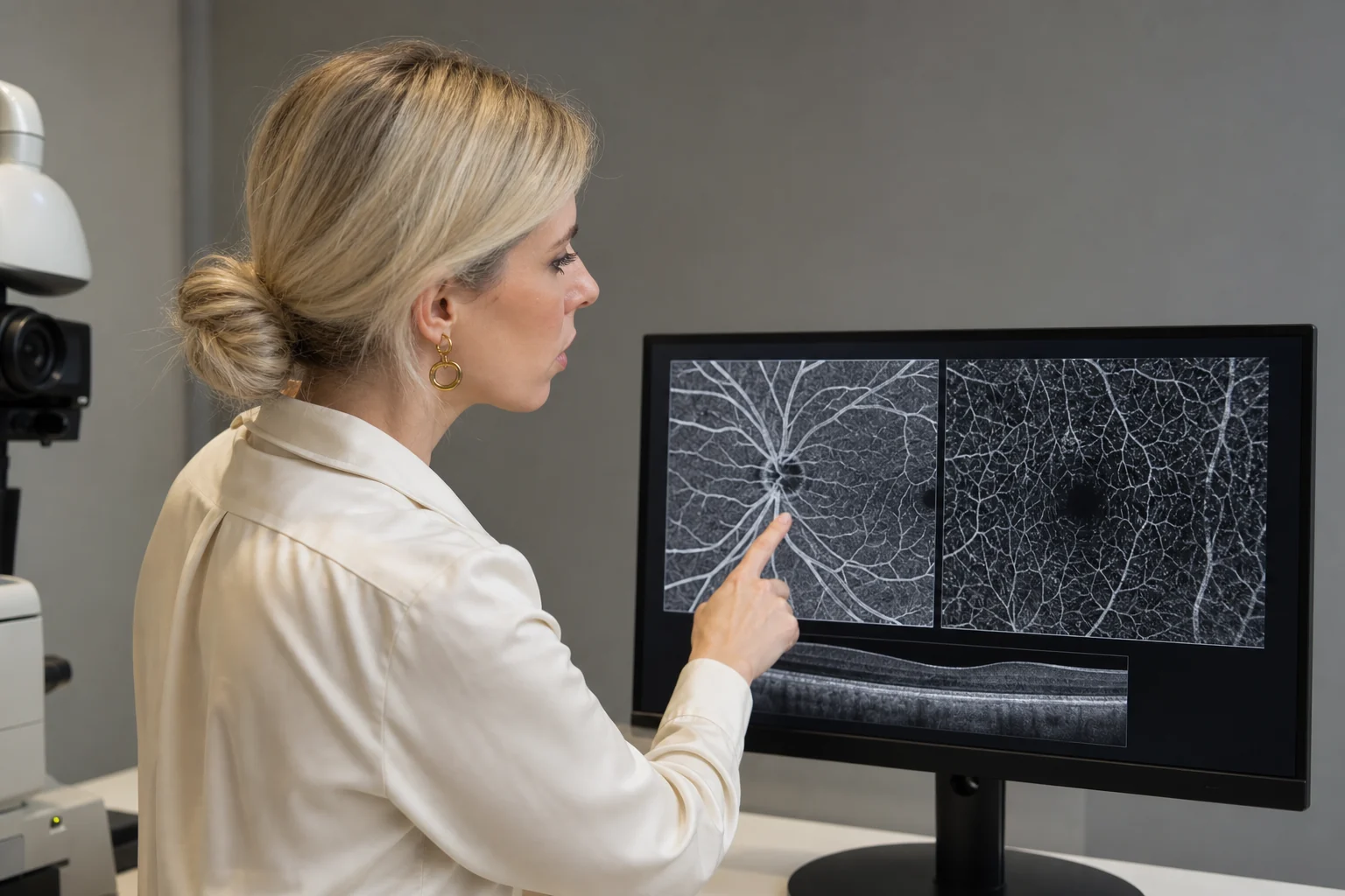
Personalised assessment
Clinical interpretation
Interpretation & limitations
Vascular mapping needs clinical context.
The doctor interprets the findings alongside structural OCT, fundus examination and the medical history.
01
Vascular network
Imaging of small vessels and the distribution of flow.
02
Without dye
Information is generated from repeated optical scans.
03
Complementary role
OCTA does not replace every other angiographic examination.
Artefacts and technical limitations can affect the image. Automated interpretation without ophthalmic assessment is not appropriate.
Frequently asked questions
Before your examination.
Concise answers about the process and preparation.
Is injected dye required?
No. OCTA does not require intravenous dye administration.
Is it the same as conventional OCT?
The process is similar, but OCTA analyses flow and complements structural OCT.
Does it replace every angiogram?
No. In some cases a different examination may be required according to the clinical question.
Vascular imaging
Is detailed mapping of the microcirculation required?
OCTA provides non-invasive vascular imaging without injected dye.
or call +30 697 421 2356
Medical information notice
This content is provided for general information and does not replace an ophthalmic examination, diagnosis or personalised medical advice.