
Optic nerve & intraocular pressure
Glaucoma & Ocular Hypertension
Progressive changes to the optic nerve that may develop without noticeable symptoms and require systematic comparison over time.
Course
Often without symptoms
Risk factor
Intraocular pressure
Monitoring
OCT & visual fields
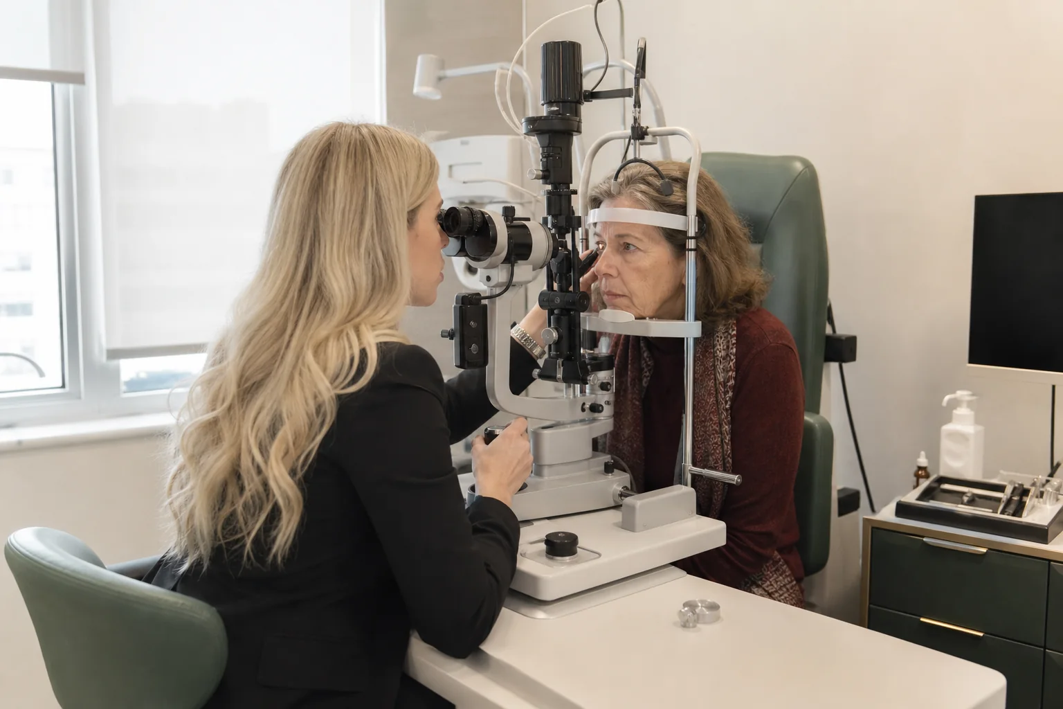
Structural assessment
Optic nerve
What is glaucoma?
A silent threat to the optic nerve.
Glaucoma is a group of conditions that cause progressive damage to the optic nerve and may lead to permanent loss of vision.
In the most common forms, there may be no noticeable symptoms in the early stages, while the peripheral vision is gradually affected.
Raised intraocular pressure is an important risk factor, but it is not synonymous with glaucoma. Diagnosis is not based on a single measurement.
High pressure may be present without established damage, while glaucomatous damage may also occur with pressure readings within the usual range.
Conditions covered
Different mechanisms, one shared aim: protecting vision.
The structure of the optic nerve, visual field function and changes over time are assessed together.
01 / OPEN ANGLE
The most common form
Primary Open-angle Glaucoma
It usually progresses gradually and without noticeable early symptoms.
- Clinical examination of the optic nerve
- OCT and visual field testing
- Comparison over time
02 / NARROW ANGLE
Anatomical obstruction
Angle-closure or Narrow-angle Glaucoma
It may develop gradually or present as an acute attack with pain, a red eye, blurred vision, haloes and nausea.
- Assessment of the anterior chamber angle
- The acute form is an emergency
- Immediate examination when symptoms occur
03 / HIGH PRESSURE
Without established damage
Ocular Hypertension
Not everyone develops glaucoma, but an assessment of overall risk is required.
- Corneal thickness
- Family history
- Personalised follow-up schedule
Symptoms
Often silent — assessment comes before symptoms.
Chronic glaucoma may progress unnoticed. An acute attack has a different presentation and is an emergency.
Could your visual field be narrowing without you noticing?
Structural and functional comparison over time is more reliable than a single measurement.
01
No early symptoms
The most common form may progress silently.
02
Change in peripheral vision
Usually noticed at more advanced stages.
03
Severe pain
When accompanied by a red eye, this may indicate an acute attack.
04
Haloes
Coloured rings around lights, accompanied by blurred vision.
05
Headache or nausea
When associated with acute eye pain, urgent assessment is required.
Severe pain, sudden blurring, a red eye, haloes or nausea? These symptoms may indicate an acute attack and require immediate examination.
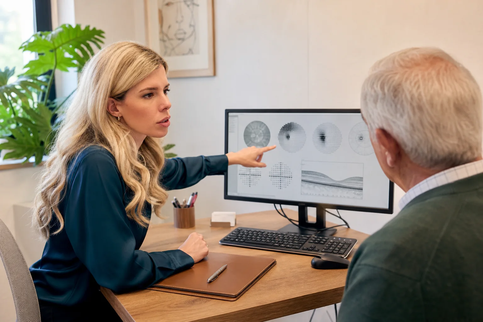
Structure & function
Comparison over time
Diagnosis & follow-up
Diagnosis requires a combination of measurements.
Glaucoma monitoring forms part of the overall assessment and is not a separate condition.
01
Tonometry
Measurement of intraocular pressure considered alongside other risk factors.
02
Gonioscopy
Assessment of the anatomy of the anterior chamber angle.
03
Optic nerve examination
Clinical assessment and photography when indicated.
04
Optic nerve OCT
Measurement of the retinal nerve fibre layer and comparison of successive examinations.
05
Visual field testing
Functional assessment of central and peripheral vision.
06
Corneal pachymetry
Adds context to the pressure measurement and overall risk assessment.
Management
The aim is to preserve the remaining vision.
Management aims to reduce the risk of further damage and is tailored to the type, stage and course of the condition.
01
Eye drops
Reduction of intraocular pressure according to the personalised plan.
02
Laser treatment
May be selected according to the type of glaucoma and the anatomy of the eye.
03
Surgery
Considered when better control of intraocular pressure is required.
04
Systematic monitoring
Repeat OCT, visual field testing and clinical examination to identify progression.
Established visual loss from glaucoma cannot usually be restored. The aim is to protect the vision that remains.
Frequently asked questions
Helpful answers.
General information that does not replace an individual medical assessment.
Does raised pressure always mean glaucoma?
No. Ocular hypertension does not always mean that damage is present, while glaucoma may also occur with pressure readings within the usual range.
Does one normal pressure reading rule out glaucoma?
No. Diagnosis is based on intraocular pressure, the optic nerve, OCT, visual fields and changes over time.
Can lost vision be restored?
Established visual loss cannot usually be restored, which is why timely diagnosis and consistent follow-up are critical.
Specialist assessment
Glaucoma may progress without noticeable symptoms.
Arrange an assessment of intraocular pressure, the optic nerve and the visual field.
or call +30 697 421 2356
Medical information notice
This content is provided for general information and does not replace an ophthalmic examination, diagnosis or personalised medical advice.