
High-resolution imaging
Optical Coherence Tomography — OCT
Detailed, non-invasive imaging of the macula and optic nerve, with objective comparison over time.
Method
Non-invasive
Imaging
Light-based
Application
Macula & optic nerve
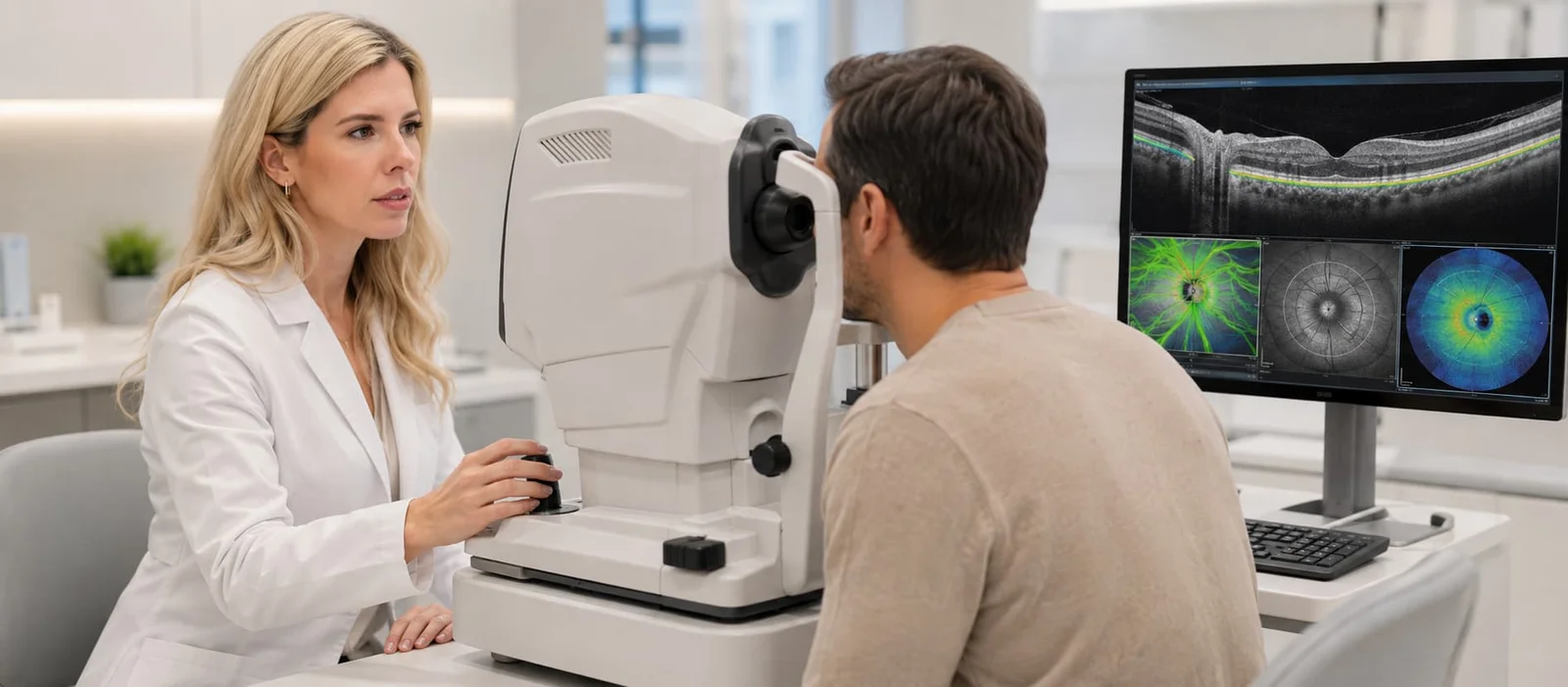
Optical Coherence Tomography
OCT
Understanding OCT
A detailed cross-sectional view of the structures of the eye.
Optical Coherence Tomography uses light to create high-resolution cross-sectional images of the retina and optic nerve.
The images reveal individual tissue layers and provide quantitative measurements that support the diagnosis and monitoring of many ophthalmic conditions.
OCT findings are always interpreted alongside symptoms, medical history and the clinical eye examination. A scan on its own does not provide an independent diagnosis.
Conventional OCT does not use radiation and does not require contrast dye. It is a different examination from OCT angiography.
What is assessed
Two areas, one precise examination.
The scanning protocol is selected according to the clinical question and the area requiring detailed assessment.
01 / OCT MACULA
Central vision
Macula
It images the layers of the macula and helps identify fluid, traction or anatomical changes.
- Age-related macular degeneration
- Diabetic macular oedema
- Epiretinal membrane
- Macular hole
- Central serous chorioretinopathy
02 / OCT NERVE
Nerve fibres
Optic nerve
It records the retinal nerve fibre layer and provides measurements that can be compared between examinations.
- Glaucoma assessment
- Investigation of suspicious findings
- Comparison of changes over time
- Additional investigation of optic nerve conditions
The process
A brief scan, without contact with the eye.
The process is straightforward and is usually completed without discomfort.
01
Positioning
You sit at the instrument and rest your chin and forehead comfortably on the supports.
02
Fixation
You look at a light target and keep your gaze as steady as possible.
03
Scanning
The instrument captures brief scans without touching the eye.
04
Assessment
The images and measurements are reviewed alongside the other clinical findings.
Will my pupils need to be dilated?
Not always. In some cases, pupil dilation may be recommended to improve image quality or to combine OCT with a comprehensive eye examination.
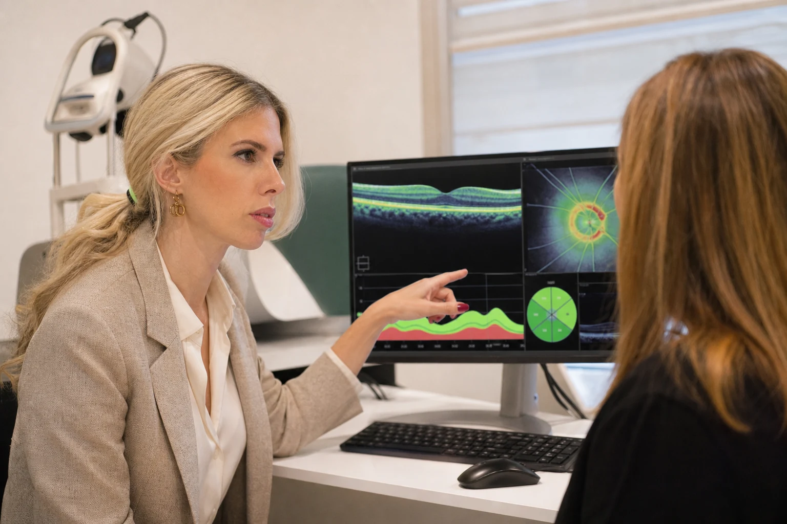
Serial measurements
Comparison over time
Findings & monitoring
Its value lies in expert interpretation.
OCT produces detailed images and measurements. They are assessed by the ophthalmologist and interpreted in the context of each patient’s clinical picture.
01
Anatomical detail
Visualisation of individual tissue layers and identification of morphological changes.
02
Quantitative measurements
Objective records that can support staging and follow-up.
03
Comparison between visits
Serial scans help assess stability or change over time.
Automated colour maps and database comparisons do not, on their own, constitute a diagnosis.
Frequently asked questions
Before your examination.
Concise answers about what to expect from the process.
Is OCT painful?
No. It is a non-invasive examination and is usually completed without discomfort or contact with the eye.
Does OCT use radiation?
No. OCT uses light to create cross-sectional images.
Is contrast dye required?
Conventional OCT does not require contrast dye. OCT angiography also does not require intravenous dye, but it is a different examination.
Why are previous scans compared?
Comparing serial scans can show whether findings remain stable or change, supporting an appropriate follow-up plan.
Detailed assessment
Do you need an assessment of the macula or optic nerve?
Arrange an ophthalmic assessment to determine whether OCT is appropriate for your clinical question.
or call +30 697 421 2356
Medical information notice
This content is provided for general information and does not replace an ophthalmic examination, diagnosis or personalised medical advice.