
Retina & macula
Macular Conditions
Conditions affecting clear, detailed central vision that require precise diagnosis, appropriate staging and personalised follow-up.
Affected area
Central vision
Symptoms
Blurring & distortion
Assessment
Fundus examination & OCT

Specialist assessment
Macula
What is the macula?
The area that enables us to see fine detail.
The macula is the central area of the retina. It provides the clear, detailed central vision used for reading, driving and recognising faces.
Macular conditions may cause blurring, distortion of straight lines, difficulty reading or a dark area in the centre of vision.
Timely diagnosis allows appropriate follow-up and, when necessary, the start of treatment. The course varies according to the particular condition and examination findings.
Some conditions may not cause noticeable symptoms in their early stages. Clinical examination and imaging are essential for an accurate assessment.
Conditions covered
Different conditions, one shared need for precise assessment.
The type, stage, symptoms and anatomical findings determine the most appropriate follow-up or management plan.
01 / AGE-RELATED CHANGE
Mainly in later life
Age-related macular degeneration
It may progress without early symptoms or cause blurring, distortion and loss of part of the central vision.
- Dry and neovascular forms
- Staging is required
- Clinical examination and imaging
02 / DIABETES
Vascular leakage
Diabetic macular oedema
Fluid leaking from retinal blood vessels causes swelling and blurring or distortion of central vision.
- Associated with diabetes mellitus
- Regular ophthalmic follow-up
- Consideration of wider health factors
03 / TRACTION
Change at the retinal surface
Epiretinal membrane
A thin layer of tissue over the macula may cause traction, wavy lines or reduced central vision.
- Assessment of symptoms
- Anatomical assessment with OCT
- Follow-up or surgical assessment
04 / OPENING
Centre of the macula
Macular hole
A small opening at the centre of the macula may cause blurring, distortion or a gap in central vision.
- Confirmation with OCT
- Staging
- Monitoring anatomical change
05 / FLUID
Beneath the macula
Central serous chorioretinopathy
A collection of fluid beneath the macula may cause blurred, distorted or darker central vision, often in one eye.
- The course varies between patients
- Duration and recurrence are important
- Management guided by imaging findings
Symptoms
Changes in central vision that require assessment.
Symptoms may be subtle or become noticeable only when each eye is checked separately.
Have you noticed a new change in your vision?
A new or sudden change requires prompt ophthalmic assessment.
01
Central blurring
Difficulty seeing faces or fine detail clearly.
02
Distortion of lines
Straight lines may appear wavy or bent.
03
Difficulty reading
Missing letters when reading or a need for more light.
04
Change in size or shape
Objects may appear different in size or shape between the two eyes.
05
Dark or blank area
A blurred, dark or blank area near the centre of vision.
06
Difference between the eyes
A noticeable difference in brightness, shape or image quality between the eyes.
New distortion or a sudden change in central vision? Do not wait for it to resolve on its own. Contact the practice promptly for an ophthalmic assessment.
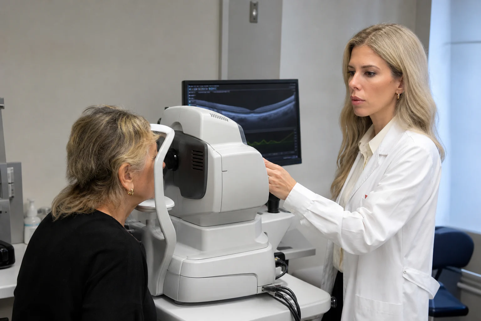
Clinical & imaging assessment
Personalised assessment
Diagnosis & follow-up
An accurate assessment requires a combination of examinations.
Assessment is tailored to the symptoms, examination findings, medical history and the particular condition.
01
Visual acuity
Assessment of central vision and comparison between the two eyes.
02
Fundus examination
Examination of the macula and retina, with pupil dilation when required.
03
Macular OCT
Detailed imaging of the retinal layers to identify fluid, traction or atrophic changes.
04
Additional imaging
Fundus photography, OCTA or other angiographic imaging when clinically indicated.
05
Comparison over time
Repeat measurements to assess progression and response over time.
Management
The plan is guided by the condition and its stage.
There is no single treatment suitable for everyone. The choice depends on the diagnosis, disease activity, symptoms and visual needs.
01
Monitoring
Regular clinical assessment and imaging to compare findings over time.
02
Intravitreal injections
A treatment option only for specific conditions and when clinically indicated.
03
Laser treatment
May be considered in selected cases, depending on the condition and examination findings.
04
Surgical assessment
Considered when anatomical change and its effect on vision warrant surgical assessment.
Monitoring, injections, laser treatment or surgery are not appropriate for every condition or every patient. The plan is chosen only after an individual ophthalmic assessment.
Frequently asked questions
Helpful answers.
General information that does not replace an individual medical assessment.
Do macular conditions affect all of your vision?
They mainly affect central vision, which is essential for reading and recognising fine detail. The effect varies according to the condition and its stage.
Can a macular condition be present without symptoms?
Yes. Some conditions or early stages may not cause immediately noticeable changes.
Why is OCT important?
OCT images the layers of the macula and helps confirm the diagnosis, establish the stage and compare findings over time.
Does every case require injections or surgery?
No. Management depends on the particular condition, its stage, symptoms and imaging findings.
Specialist assessment
Have you noticed blurring or distortion in the centre of your vision?
Arrange a specialist macular assessment and receive a personalised diagnostic and follow-up plan.
or call +30 697 421 2356
Medical information notice
This content is provided for general information and does not replace an ophthalmic examination, diagnosis or personalised medical advice.