
Retinal photography
An objective record of the appearance of the fundus.
Digital documentation of the retina, macula, retinal vessels and optic nerve.
Record
Digital image
Value
Comparison over time
Role
Complementary
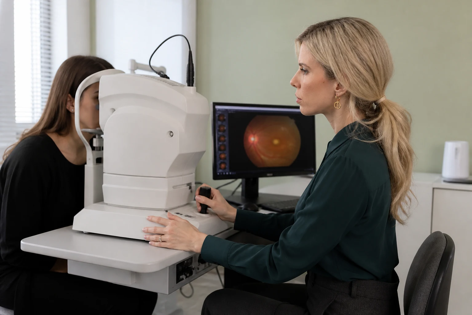
Eye examinations
Fundus Photography
Documentation & follow-up
An image becomes a reference point for monitoring change.
Digital fundus photography creates an objective photographic record of the posterior structures of the eye.
Images can be used to document, compare and monitor changes between visits.
Photography supports clinical examination but does not always replace fundus examination or capture every peripheral area in the same way.
Whether images can be obtained without pupil dilation depends on the actual equipment and conditions. The doctor decides whether dilation and a full fundus examination are required.
Clinical uses
Documented follow-up, one image compared with the next.
Photographic records allow objective comparison of findings.
01 / RECORD
Retina
Fundus findings
Documentation of central findings for the clinical record.
- Diabetic retinopathy
- Vascular changes
- Retinal conditions
- Macula
02 / COMPARISON
Optic nerve & vessels
Objective record
Comparison of appearance between visits.
- Optic nerve appearance
- Retinal vasculature
- Sequential images
- Documentation of change
The process
A brief image with precise fixation.
Image quality is checked before the process is completed.
01
Positioning
The chin and forehead are supported on the device.
02
Fixation
The gaze remains on a specific target.
03
Image capture
The brief photograph may be accompanied by a bright flash.
04
Quality check
Additional images may be taken for other areas or improved clarity.
Is pupil dilation required?
Not always, when the equipment and conditions allow. The doctor makes this decision and a full fundus examination may still be required.
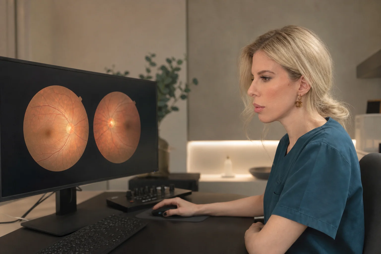
Personalised assessment
Clinical interpretation
Findings
The value lies in comparison and accurate interpretation.
The ophthalmologist reviews the images alongside the medical history and clinical examination.
01
Documentation
An objective record of the appearance of the fundus.
02
Comparison
Identification of possible changes between visits.
03
Complementary testing
Combination with fundus examination, OCT or another test when indicated.
Photography does not always replace direct clinical examination, and results must not be interpreted automatically.
Frequently asked questions
Before your examination.
Concise answers about the process and preparation.
Is image capture painful?
No. It is brief, although a bright flash may be noticed.
Is it always performed without dilation?
No. This depends on the equipment, the pupil and image quality.
Does it replace fundus examination?
Not always. The doctor decides whether direct clinical examination and pupil dilation are required.
Objective documentation
Do you need documentation or comparison of fundus findings?
Digital fundus photography supports documented follow-up.
or call +30 697 421 2356
Medical information notice
This content is provided for general information and does not replace an ophthalmic examination, diagnosis or personalised medical advice.