
Retina & optic nerve
A detailed examination of the inside of the eye.
Fundus examination allows assessment of the macula, retina, retinal vessels and optic nerve.
Examination
Clinical
Area
Posterior segment
Dilation
When required
Eye examinations
Fundus Examination
What is fundus examination?
A direct view of the structures that support vision.
Fundus examination is a core investigation for diagnosing and monitoring many eye conditions.
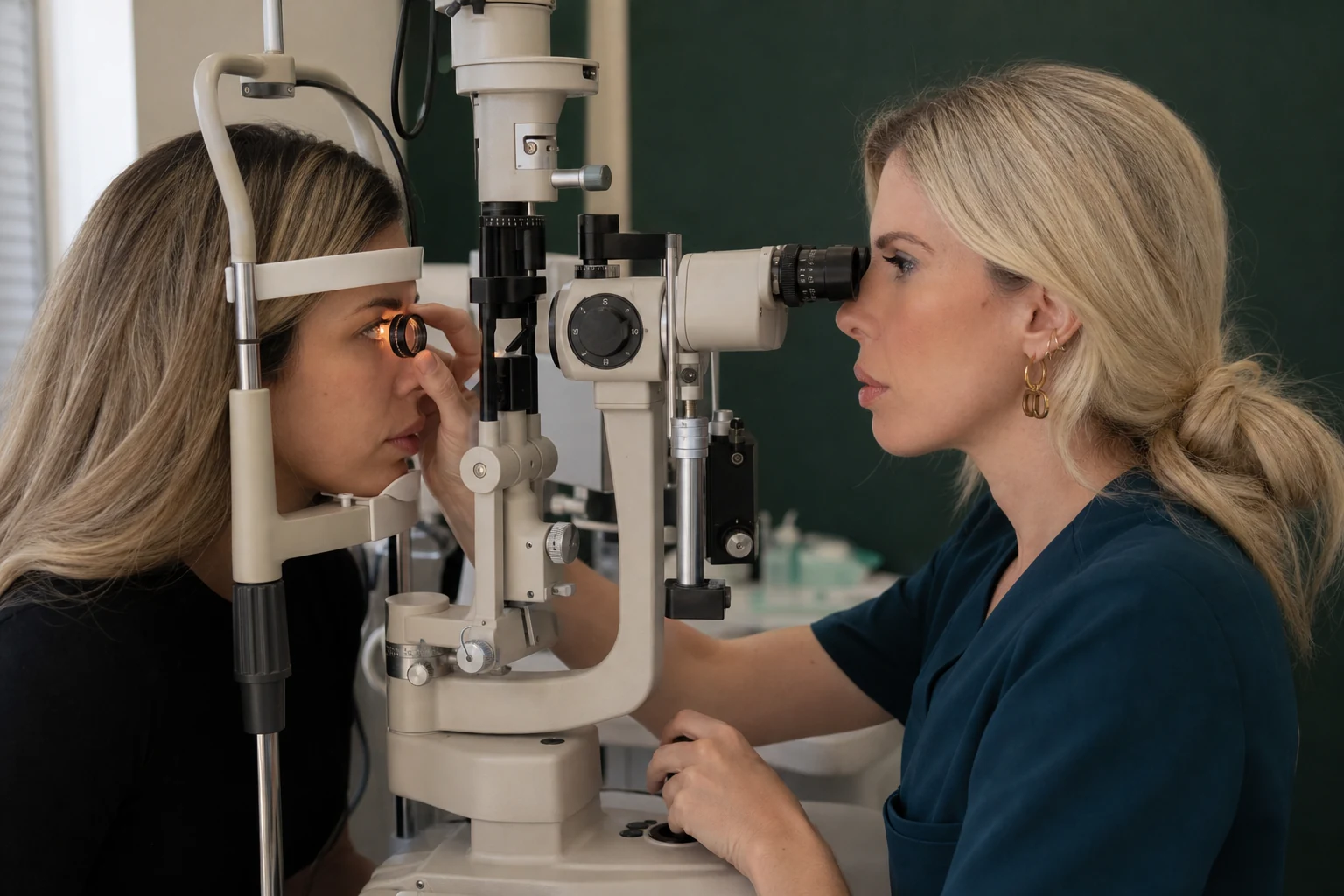
It may be performed at the slit lamp using specialist lenses or with other appropriate techniques.
In many cases, drops are used to dilate the pupil so that the peripheral retina can be examined in greater detail.
The new onset of numerous floaters, flashes or a shadow in the visual field requires urgent ophthalmic assessment.
What it can assess
From the macula to the peripheral retina.
The clinical findings guide the need for additional imaging.
01 / RETINA
Central & peripheral vision
Retina & macula
Assessment of changes that may affect central or peripheral vision.
- Diabetic retinal changes
- Macular conditions
- Retinal tears or detachment
- Retinal vascular occlusions
02 / NERVE
Vessels & neural structures
Optic nerve & vitreous
Assessment of the optic nerve head and changes within the vitreous.
- Optic nerve appearance
- Vitreous changes
- Inflammation
- Haemorrhage or other findings
The process
A detailed clinical examination with or without pupil dilation.
The technique is selected according to the area that needs assessment.
01
Initial assessment
Discussion of symptoms and assessment of vision.
02
Pupil dilation
Drops are used when a clearer view of the peripheral retina is required.
03
Fundus examination
Assessment using suitable lenses and illumination.
04
Conclusion
Interpretation of findings and a decision about imaging or follow-up.
After pupil dilation
Near vision may be temporarily blurred and the eyes more sensitive to light. Bring sunglasses and follow the doctor’s individual guidance.

Personalised assessment
Clinical interpretation
When it is recommended
Symptoms and history determine the scope of the examination.
Fundus examination is used both for urgent symptoms and for monitoring known conditions.
01
New symptoms
Floaters, flashes, a shadow, or reduced or distorted vision.
02
Systemic history
Diabetes mellitus or conditions that may affect the retina.
03
Follow-up
A known retinal or optic nerve condition, or related pre- or post-operative assessment.
The duration of the effects of pupil dilation varies. The doctor provides individual guidance.
Frequently asked questions
Before your examination.
Concise answers about the process and preparation.
Are eye drops always needed?
No. Pupil dilation is used when a more detailed examination is required, particularly of the peripheral retina.
What might I experience afterwards?
Temporary near blur and increased sensitivity to light. The duration varies between patients.
Is fundus photography the same examination?
No. A digital image documents findings but does not always replace direct clinical examination.
Prompt assessment
Have you noticed new floaters, flashes or a change in vision?
Fundus examination supports prompt assessment of the retina.
or call +30 697 421 2356
Medical information notice
This content is provided for general information and does not replace an ophthalmic examination, diagnosis or personalised medical advice.