
Central & peripheral vision
Mapping the space you can perceive.
Functional mapping of central and peripheral vision for diagnosis and follow-up.
Type
Functional test
Assessment
Each eye separately
Requirement
Concentration
Eye examinations
Visual Field Testing
What visual fields assess
Vision extends beyond the point at which we look.
Visual field testing records how well the space around and ahead of the central point of fixation is perceived.
It assesses central and peripheral visual function and is used primarily to diagnose and monitor glaucoma.
It can also support the investigation of optic nerve conditions and selected neurological disorders.
The first test can be influenced by familiarity with the process. The doctor reviews reliability indices and may recommend a repeat test.
When it is used
Functional measurement for consistent follow-up.
A single result is not always sufficient; change is often assessed through a series of tests.
01 / GLAUCOMA
Diagnosis & progression
Glaucoma assessment
Detection and monitoring of functional visual field defects.
- Initial assessment
- Ocular hypertension
- Suspicious optic nerve appearance
- Response to treatment
02 / NERVE
Neuro-ophthalmology
Optic nerve & visual pathways
Investigation of unexplained defects and known damage.
- Optic nerve conditions
- Neurological disorders
- Unexplained defects
- Comparison over time
The process
Steady fixation and a simple button.
The test is not painful but requires concentration and cooperation.
01
Positioning
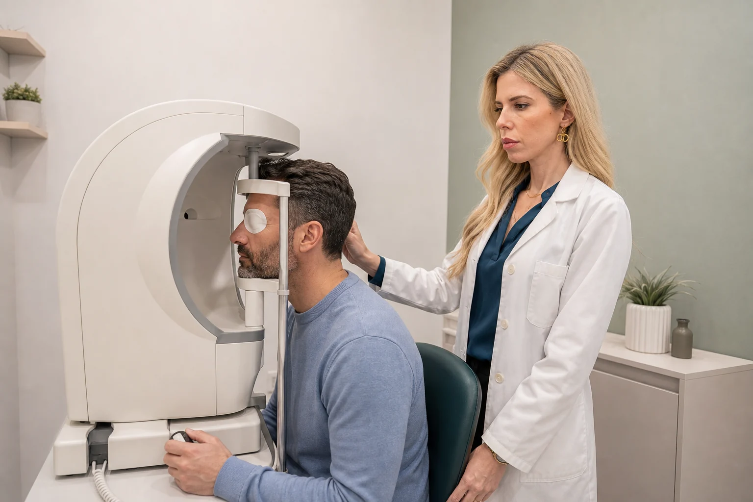
One eye is covered while the other looks into the bowl.
02
Fixation
The gaze remains steady on the central target.
03
Response
Press the button whenever you perceive a light stimulus.
04
Second eye
The other eye is tested separately and reliability indices are checked.
Before the test
Bring your glasses, rest beforehand if possible and ask any questions before starting. You are not expected to see every light.

Personalised assessment
Clinical interpretation
Reliability & repetition
The consistency of responses determines the value of the result.
Fatigue, loss of fixation, incorrect responses or blurred correction can affect reliability.
01
Reliability indices
The doctor checks whether the result can be interpreted safely.
02
Learning effect
Familiarity often improves test quality on a subsequent occasion.
03
Comparison of tests
Multiple measurements help distinguish genuine change.
Do not worry if you do not see every light stimulus; some are intentionally very faint.
Frequently asked questions
Before your examination.
Concise answers about the process and preparation.
Is the test painful?
No. It only requires fixation and pressing a button when you perceive a light stimulus.
Why might it need to be repeated?
Fatigue and familiarity affect reliability, particularly during a first test.
Are both eyes tested together?
No. Each eye is tested separately.
Functional assessment
Do you need an assessment of peripheral vision?
Visual field testing is central to monitoring glaucoma and optic nerve conditions.
or call +30 697 421 2356
Medical information notice
This content is provided for general information and does not replace an ophthalmic examination, diagnosis or personalised medical advice.